
Gore donates new 3D X-ray imaging system to UD
Some people collect classic cars. Others stockpile antiques.
University of Delaware’s Gerald (Jerry) Poirier acquires complex analytical tools — the kind that enable the intricate and sophisticated research of engineers, art conservationists, physicists and more.
Poirier, who manages the Advanced Materials Characterization Lab in UD’s Patrick T. Harker Interdisciplinary Science and Engineering Laboratory, recently had the opportunity to cross off an item on his wish list: UD’s first 3D X-ray imaging system.
The new system includes a micro-CT system and a nano-CT system that will enable researchers to scan through solid objects, such as the human body or the earth, for an unprecedented look at the object’s internal structure. The micro-CT system is a high-speed X-ray microscope that uses a form of computed tomography (CT) to create 3D images. Tomography is a technique used to scan solid objects in slices or sections and record them as a series of pictures that can be viewed individually (two-dimensional) or as a whole (three-dimensional).
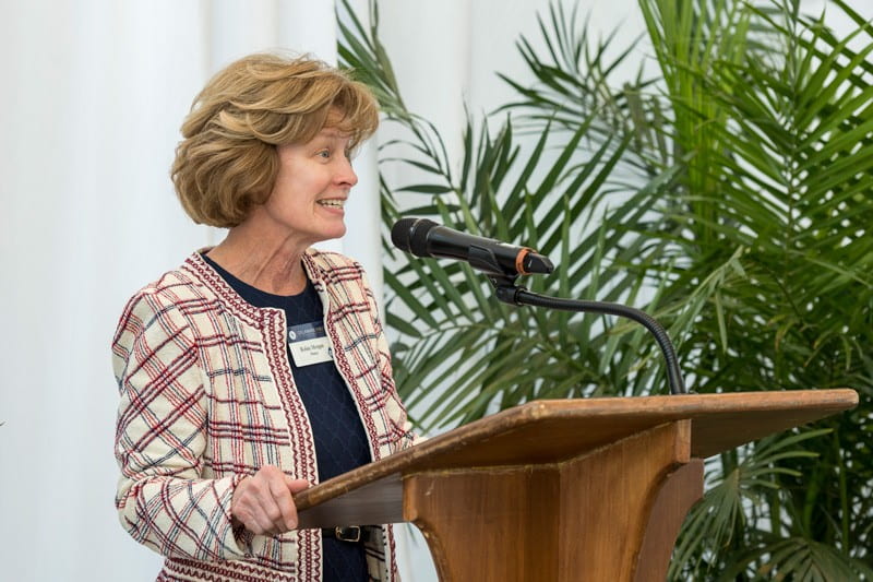
UD Provost Robin Morgan explains how a new 3D X-ray imaging system, made possible through a gift from W. L. Gore & Associates, Inc., will help elevate the University’s research competitiveness.
The nano-CT system is extremely fast and capable of delivering high-resolution, 4D images — it is geared toward looking at sub-micron-size structures, smaller than the diameter of a human hair, such as particles and lower-density, soft materials like those found in carbon deposits, for example.
UD leaders, researchers, students and community members gathered in the Harker ISE Lab atrium to celebrate the arrival of the new imaging system, designed to enhance UD’s research competitiveness, on Wednesday, March 27. The new instrumentation was made possible through a gift from W. L. Gore & Associates, Inc., a long standing partner with UD to advance research and discovery.
“We had a vision when this facility was built, that Harker Lab would promote the interdisciplinary work of our faculty and students, while creating opportunities for collaborative research with members of the broader community, industry and other partners,” UD Provost Robin Morgan said in opening remarks. “These two instruments take us a step further toward realizing that vision and having this building be everything that we imagined. We can’t do these things without the kind of help that Gore has provided, and we are very grateful for this investment.”
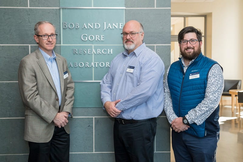
Gore’s Greg Hannon (left), Jeffrey Ledford (center) and John Delluca (UD alumnus) stand outside the Bob and Jane Gore Research Laboratory. W. L. Gore & Associates, Inc., a long standing partner with UD to advance research and discovery, donated funds for UD’s first 3D X-ray imaging system, which is located in the Advanced Materials Characterization Lab.
Greg Hannon, Gore global technology leader, also spoke of the long-standing connection between Gore and UD.
“We are confident that the team here at UD will make good use of the new 3D X-ray imaging system and we look forward to hearing more about your discoveries,” Hannon said.
Advanced equipment right on campus
The new instrumentation will help researchers address wide-ranging studies in material and life sciences.
UD doctoral student Angela Cuadros said the new equipment will enable her to run characterization studies necessary to complete her doctoral degree. Cuadros is developing low-cost ways to enhance X-ray image processing for medical and security applications, under the advisement of Gonzalo Arce, Charles Black Evans Professor of Electrical Engineering. Cuadros is focused on methods to create colored X-rays that can illuminate more than how dense an object is (like gray-scale X-rays), but also tell researchers something about the object’s material and chemical properties.
To facilitate Cuadros’s work, the instrument manufacturer (Rigaku) built an attachment for the micro-CT X-ray machine to hold specialized filters and grids, known as color-coded apertures, to filter various wavelengths of electromagnetic radiation. Cuadros said having the ability to process images at multiple energy levels, or wavelengths, known as spectral imaging, can provide an additional layer of information.
“Spectral tomography allows for ‘colored’ rays, rather than the traditional gray-scale X-rays most of us are familiar with,” she said. “In tissue, the color describes characteristics, such as healthy or abnormal. It also would help distinguish non-harmful liquids like water and soft drinks from those that are highly flammable, such as gasoline, or explosive, such as hydrogen peroxide, among other things.”
For soil scientist Yan Jin, having these sophisticated instruments located just downstairs from her office on the Harker Lab’s fourth floor is an exciting development. Jin is collaborating with colleague Harsh Bais, both professors in UD’s Department of Plant and Soil Sciences, to understand how a UD-developed beneficial microbe named UD1022 increases water retention in soil to mitigate drought.
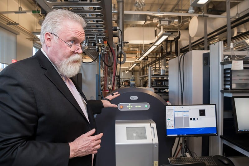
Thomas McNulty, senior vice president and general manager at Rigaku Americas Corporation, discusses the capabilities of UD’s new 3D X-ray imaging system.
For the last four years, Jin and her students have traveled to the National Institute of Standards and Technology (NIST) in Gaithersburg, Maryland, a few times per year to run experiments using NIST’s high-powered neutron imaging and 3D X-ray tomography instruments. Having some of the same equipment available right on campus will allow Jin and her students to ask new questions, run additional experiments and keep the research moving forward in the downtime between trips to NIST.
“Now that this 3D X-ray system is available right here at UD, I can sign up for instrument time online and be more open-minded about the experiments our group might want to work on,” said Jin.
According to Poirier, few other academic institutions offer the top-notch facilities and equipment found in Harker Lab, and UD is helping set the standard among its peer institutions. The 3D X-ray imaging system is a technology that is in demand, too.
“Seeing is believing. When you can scan through something in 3D, rotate it around and manipulate it, that’s quite extraordinary. This is a technology the University needed,” said Poirier. Arce and Ken Barner, chair of the Department of Electrical and Computer Engineering, worked with Poirier on the acquisition.
Advanced Materials Characterization Lab
The Advanced Materials Characterization Laboratory in Harker ISE Lab opened in 2014. Today, the facility accommodates sophisticated instruments and equipment that allow researchers to complete 28 different scientific techniques. Managed by Poirier, the AMCL accommodates the research needs of 500 users from UD and approximately 50 external companies. The facility also has two lab coordinators Jing Qu and Chin-Chen Kuo. Poirier, Qu and Kuo all are UD alumni.
The instruments are available to all members of the University community, as well as local researchers and companies. Local and international users can make use of the equipment remotely, too, through sample shipment and data transfer.
A short course on how to use the new CT X-ray machines is currently in development, with the first offering for students coming later this spring. Additional short courses are available on each of the facility’s other instruments. For more information, contact Jerry Poirier via email gpoirier@udel.edu or by phone at 302-831-6795.
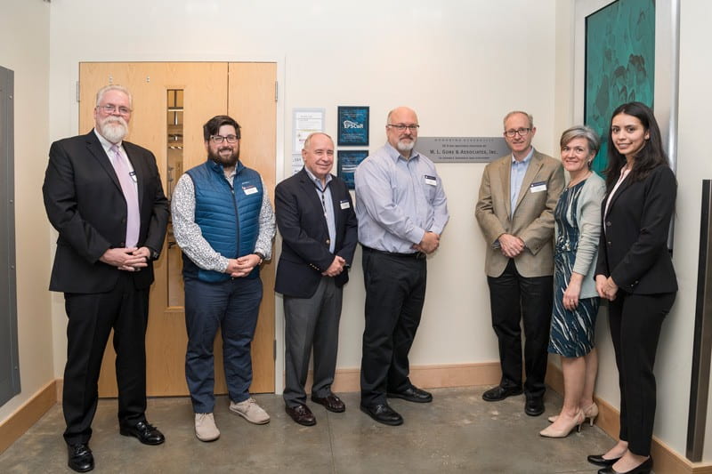
Representatives from Gore, Rigaku and UD pause before touring the Advanced Materials Characterization Laboratory. Pictured from the left: Tom McNulty (Rigaku), Gore representatives John Delluca (a UD alumnus), Michael Dougherty, Jeffrey Ledford, Greg Hannon, Amy Calhoun, and UD doctoral student Angela Cuadros.